

 Global
  United States
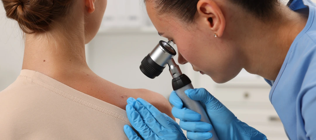
What is AI Skin Cancer Detection?
Melanoma is a type of skin-based cancer like squamous cell carcinoma and basal cell carcinoma that originates from melanocytes, which are basically the cells that produce a pigment called melanin responsible for coloration of the skin. It metastasizes and invades surrounding tissues and distant organs through uncontrolled cellular proliferation, unlike benign melanoma lesions. Although melanoma is categorized as one of the most aggressive forms of skin cancer due to its metastatic potential, it has a higher treatment success rate with early diagnosis.
Not to mention, medtech solutions, such as visual assessment alone, may not be sufficient or consistent enough to differentiate benign nevi, seborrheic keratosis, or atypical moles from cancerous cells during visual screening. As Artificial Intelligence (AI) is stepping into every domain of the modern era, it has also taken up the challenge of earliest melanoma diagnosis. AI skin cancer detection tools are now integrated in mobile or web applications and diagnostic machines or platforms that earlier had low specificity and were limited by hardware fixtures across healthcare facilities. This accelerates skin lesion image AI pattern recognition, analysis, and medical assessments, leading to more accurate and faster treatments.
Apart from this, another global challenge is the increasing volume of patients as compared to available dermatologists, as traditional methodologies involve in-depth visual examination or comparison with benign lesions, biopsy, and histopathology. Computer Vision (CV), Deep Learning (DL – a subset of Machine Learning), and AI development services for medical imaging and derma interpretation can enhance clinical diagnostic pathways. These technological advancements predict melanoma risk by analyzing multispectral scans, clinical visuals, lesion, skin, and dermoscopic images captured through apps and machines accurately.
Given below are a few market-related facts around skin cancer detection.
- In 2025, the worldwide market for skin cancer detection devices had reported a value of US $1,073.8 million, which is projected to reach an approximate value of US $3,133.8 million by 2035, surging at a CAGR of 11.3% during this forecast period.
- In 2024, the market for skin cancer diagnostics was valued at US $9.58 billion, which is forecasted to surge at a CAGR of 5.7% to reach around US $15.67 billion by 2033.
- As of 2026, the global market value for AI in dermatology has been reported to be around US $9.32 billion and may increase at a CAGR of 15.43% to reach approximately US $19.09 billion by 2031.
- The global AI skin analysis market has been reportedly valued at US $2.13 billion in 2026 and may increase up to US $6.3 billion in 2033, rising at a CAGR of 16.8% during this period.
- The market size of AI skin analysis instruments was valued at around US $2.3 billion as of 2024 and is expected to surge to reach a value of US $8.5 billion by 2033, rising at a CAGR of 14.1%.
In this blog, we will delve into the intricacies of AI for medical imaging, especially for cancerous skin lesions and melanoma enabled with computer vision in healthcare, and DL-driven next-gen diagnostic advancements for improving survival rates.
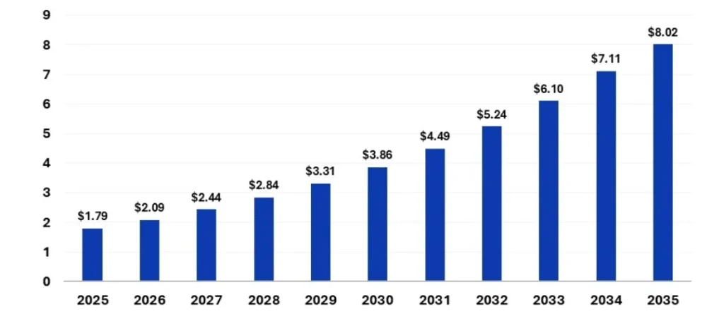
Source: Precedence Research
Growing market size of AI melanoma detection during the forecast period 2025 to 2035
How does AI Skin Cancer Detection Work?
AI algorithms, such as multi-layer Convolutional Neural Networks (CNN – a DL, for example, ResNet, U-Net, Attention U-Net, DeepLabV3+, Mask R-CNN, DenseNet, EfficientNet and Vision Transformers) are trained on huge datasets of mole and lesion images to precisely detect melanoma by learning visual symptoms caused by the presence and activities of malignant cells. Their architectures learn from pixel-level representations that are extracted directly from annotated masks, producing a binary segmentation mask, thus identifying precise lesion boundaries.
These types of unsupervised learning ML models can be flexibly deployed through AI model deployment techniques over diagnostic apps, tools, machines, platforms, clinical and telemedicine settings to analyze patient images for skin cancer detection and have been proven to generate accurate outcomes and specificity. Given below is a typical structured workflow involving multiple stages of AI-enabled melanoma detection systems.
Visual Data Acquisition
Smartphones, embedded medical scanners, devices, and portable dermatoscopes in hospitals, dermatology clinics, rural healthcare stations, point-of-sale centers, research institutions, clinical trial data analysis, and telemedicine platforms are used to capture high-resolution images for further analysis. The large-scale datasets, which may also be created using retrieval-augmented generation, contain annotated or data-labelled images of various categories of lesions, moles, skin types, histopathological confirmations, Optical Coherence Tomography (OCT), and Reflectance Confocal Microscopy (RCM) images, for example, ISIC, HAM10000, PH2, BCN20000, etc., datasets.
Data Preprocessing
The system enhances raw captured images in terms of the following.
- Image Resizing: Common standards for different types of neural networks are 224×224, 299×299, and 512×512 pixels, followed by orientation modification.
- Color Normalization: Consistent illumination, contrast, and generalization.
- Occlusion Removal: Hair removal, morphological filtering, inpainting, and DullRazor techniques for melanoma cancer detection.
- Lesion Segmentation: DL-based lesion region classification by separating lesion pixels from healthy skin pixels as compared to traditional methods of thresholding, edge detection, region growing, and active contours for enhanced feature extraction.
- Noise Reduction: Gaussian, median, and bilateral filtering to preserve lesion boundaries, image quality, and improve model performance.
- Data Augmentation: At times, medical datasets remain limited to consumer images resulting in less generalization and overfitting. This can be overcome using synthetic variations (data augmentation, rare lesion generation, and training dataset balancing) to improve robustness (rotation, flip, scale, crop, color jitter, and contrast modification using methods like MixUp, CutMix, GANs, diffusion models, variational autoencoders, etc.).
Pattern Extraction
Post-segmentation, the system extracts required characteristics from the images and applies the ABCDE rule of distinguishing between the benign and cancerous lesions. AI melanoma detection compares asymmetry index, border irregularity, color variations (mean color values and pigment distribution), circularity or compactness (diameter greater than 6 mm), and evolving appearance (texture as per Gray-Level Co-occurrence Matrix, local binary patterns, and fractal dimensions). The extracted features are fed into ML classifiers, including Support Vector Machines (SVM), Random Forests, Logistic Regression, etc.
Feature Learning
Deep neural networks showcase automatic learning of hierarchical image features through melanoma pattern detection directly from the data in lieu of manual feature definition. Here is the level-wise representation used by them for better diagnostic performance.
- Low-Level Features: These are learned by the network’s early layers, which include edges, lines, and color gradients, similar to fetus characterization in assisted reproduction treatment.
- Mid-Level Features: These include pigment structures, texture variations, and vascular patterns that are learned by the intermediate layers of the neural network.
- Higher-Level Features: The deeper layers of the network recognize malignant morphology, melanoma-specific signatures, and complex lesion structures.
The trained models undergo parametric upgrade and gradient-based optimization when developed through AI integration services using common optimizers, such as Adam, AdamW, SGD with Momentum, and RMSprop.
Threat Evaluation
The system utilizes parametric metrics to evaluate sensitivity (needs to be low for better melanoma detection rate), specificity (needs to be high for correct identification of benign lesions), accuracy, Area Under the Receiver Operating Characteristic Curve (AUC-ROC that discriminates capability needs to be above 0.90), and F1 score (for imbalanced datasets and recall).
To reduce prediction errors in melanoma cancer detection, loss functions are used, such as binary cross-entropy (melanoma versus non-melanoma classification), categorical cross-entropy (multi-class lesion classification), and focal loss (class imbalance addressal). As per the training, the system assigns a malignancy score that depicts the probable chances of melanoma or any other type of skin cancer.
Medical Advisory Support
The system then cites previous case studies and findings that assist dermatologists and pathologists in performing informed decision-making, diagnostic steps, and further referrals, such as in the case of AI breast cancer detection. Explainable AI improves interpretability of the model’s decision-making process and includes the following methods.
- Grad-CAM: Gradient-weighted Class Activation Mapping that highlights image regions for better predictions. The heatmap showcases areas attracting the model’s attention, AI medical diagnostics hotspots, and potential malignancy indicators.
- Saliency Maps: These maps rely on pixels and validate whether clinically relevant structures are focused upon.
- SHAP Analysis: Shapley Additive Explanation values quantify contributions of features to the model’s results to provide transparency into regulations in healthcare and clinical acceptance.
Furthermore, multimodal AI skin cancer diagnostic systems powered by agentic AI solutions and generative AI services extract patient metadata, including their age, sex, skin type, family history, lesion location, gender, etc., to combine visual and clinical information and improve diagnostics. Multimodal large language model and transformer models integrate heterogeneous data sources into a unified framework that generates predictions.
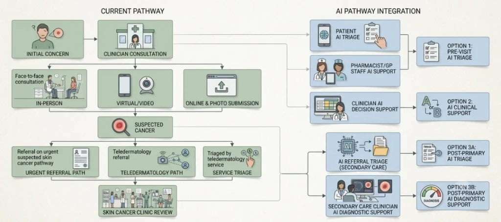
Diagram depicting inclusion of AI melanoma detection in regular clinical diagnostic pathway
KritiKal: Breaking Barriers in AI Skin Cancer Diagnostic Solutions
In this blog, we discussed how AI is enhancing melanoma detection through lesion localization, feature extraction, and segmentation within the images obtained from embedded medical devices, wearable health monitoring devices, and smartphone applications. DL-based CNNs and transformer models support early AI skin cancer diagnostic by generating precise lesion boundaries and detecting suspicious skin alterations prior to the appearance of visible symptoms.
Not only does AI improve accessibility and analysis through such medical imaging techniques, but it also limits unnecessary biopsies. This assists clinicians and pathologists to focus on decision-making, prognosis, risk-wise case prioritization, healthcare cost reduction, and treatment planning through therapeutic medical equipment. Nowadays, AI is deeply rooted in clinical workflows across industries, such as teledermatology, NHS trials, triage systems, and more.
KritiKal Solutions can uplift your current derma analytics performance to the next level by overcoming common challenges in this sector, including dataset bias, privacy concerns, regulatory approvals, variable image quality, false diagnoses risks, model updates, and overreliance. Our state-of-the-art multimodal AI systems, vision transformer, combine images and patient data to generate apt diagnostics alongside model explainability, diverse datasets, federated learning, personalized safety assessments, performance benchmarking, clinical validation, increased specificity, sensitivity, and accuracy.
We understand that future dermatology is likely to rely on human-AI collaborative oversight of skin cancer detection that would improve overall patient outcomes. Please get in touch with us at sales@kritikalsolutions.com to know more about our medtech, AI, and vision-based products, platforms, services, and realize your business requirements.

Gaurav Singh currently works as an AI/ML Engineer at KritiKal Solutions. He is proficiently skilled in developing and deploying solutions based on generative AI, agentic AI, computer vision, and LLMs. With his ability to work efficiently in teams, he has assisted KritiKal in delivering various projects to some major clients.