

 Global
  United States
Breast cancer is the most common type of cancer after skin and lung cancers and is the leading cause of cancer-related deaths among women worldwide. In this type of disease, abnormal cells grow out of control to form tumors and even become malignant in the coming stages due to malfunctioning of tumor suppressor genes BRCA1 and BRCA2 that empower growth inhibitors. Additionally, genetic mutations in PTEN, TP53, STK11 and CDH1 are also commonly observed in such patients. Certain key biomarkers on cancer cells including ER, PR, HER2 and Ki-67 indicate cancer management by determining the subtype and growth rate of breast cancer. About 1.3 million women are diagnosed on a yearly basis with breast cancer. The most common techniques for accurate and early breast cancer diagnostics include mammography, ultrasound and magnetic resonance imaging (MRI), which play a pivotal role in strategizing and mediating prognosis and treatment plan.
In certain cases, these techniques may provide false negative or false positive results and require additional biopsies, expert consultation and interpretation. Such limitations can be overcome with Artificial Intelligence (AI) which offers a transformative approach to the working of these imaging techniques. It supports enhanced breast cancer detection by leveraging computational power for accuracy and reducing human error and lowers healthcare costs effectively. The breast cancer imaging market in the United States was valued around US $1.7 billion in 2019 and is forecasted to reach US $2.4 billion by 2026. Let us further explore various imaging modalities, algorithms, processing techniques and related challenges of AI breast cancer detection.
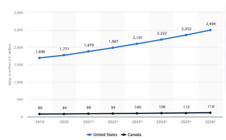
Projected global market value of AI-based breast cancer diagnostics from 2019 to 2026
Artificial Intelligence in Mammography
AI has been seen to improve imaging outcomes, avoid aggressive treatment plans for patients and save lives over the years. It has thoroughly enhanced the working of mammography, the most commonly used effective imaging tool for cancer diagnostics and screening which can accurately detect the smallest of lesions, lumps, masses, calcifications, asymmetries, tissue abnormalities, distortions contained in breast and other symptoms. AI can improvise screening, pathology, diagnostics and data analysis in clinical research areas. We are now moving forward to Machine Learning and Deep Learning algorithms involved in AI breast cancer detection.
Supervised Learning
This type of learning involves training of the ML model on labeled datasets and known outcomes that are presence or absence of cancer.
- Random Forests: These are ensemble learning methods built by aggregating multiple decision trees for prediction. They can interpret the features that form the main factors for prediction by handling high-dimensional data and feature-wise metrics.
- Support Vector Machines: SVMs are one of the types of supervised learning which are mainly used for classifying extracted features as the AI reading mammograms progresses. These features can be tumor shape, size, texture and help in classifying the cancer as benign or malignant, based on a hyperplane separating the data calculated by the algorithm.
- Convolutional Neural Networks: CNNs can decipher visual data by capturing spatial hierarchies and are well-suited for image-based tasks. With multiple layers such as convolutional, pooling and fully connected layers, they can automatically learn features as the process involves AI reading mammograms, to distinguish between benign and malignant lesions. Techniques like transfer learning where models are pre-trained thoroughly with large data sets of mammographic images can be used for better performance.
Unsupervised Learning
These types of models do not rely on labeled data and extract patterns and groupings within the provided data.
- Clustering: It includes techniques like k-means and hierarchical clustering that can identify subtypes of breast lesions as per imaging features correlated with varied biological behaviors or treatment responses.
- Dimensionality Reduction: It includes techniques such as Principal Component Analysis (PCA) and t-Distributed Stochastic Neighbor Embedding (t-SNE) that can reduce complexity of imaging for easy visualization and analysis.
Deep Learning
Deep Learning (DL) is basically a subset of machine learning which utilizes different types of neural networks that have multiple layers, also known as deep neural networks like Recurrent Neural Networks (RNNs), MIRAI, AlexNet etc. that are used in the application of artificial intelligence in mammography.
- Deep CNNs: DCNNs can handle large-scale imaging data and are adept at learning hierarchical representations of features. They identify subtle patterns directly from pixel values in the mammographic images and improve lesion detection accuracy.
- Generative Adversarial Networks: GANs are composed of a generator and discriminator network and can be used for data augmentation, which is creation of synthetic mammographic images for augmenting training datasets in case labeled dataset is not available or is scarce.
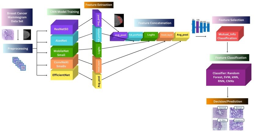
CNN Architecture for enhanced breast cancer detection
Data Processing
- Data Preprocessing: It is necessary for apt performance of AI breast cancer detection. It includes normalization where image pixel values are scaled to a common range for ensuring a uniform input to the AI model. It also involves data augmentation techniques like rotation, flip, zoom and artificial expansion of training dataset for model generalization. Another important technique in this segmentation which isolates regions of interest or tumors from the tissue in background in mammographic images to reduce noise and assist the model to focus on relevant features.
- Feature Extraction: DL models employ feature extraction for identifying and quantifying attributes that indicate breast cancer from imaging data. This can include texture analysis that quantifies patterns in pixel intensities in images to differentiate between various types of tissues and lesions. And shape analysis where geometry of lesions is analyzed for irregularities that indicate malignancy. DL models can also extract histopathological features such as cellular morphology from biopsy images for breast cancer diagnostics.
Integration with Imaging Modalities
The aforementioned ML and DL models can easily integrate with various imaging modalities such as mammography which involves low dose X-rays projected between imaging plates, sonography that is enabled with sound wave produced by transducers to differentiate between fluid-filled cysts and solid mass, or MRI that utilizes magnets and radio waves for 3D imaging.
- Mammography: AI breast cancer detection enables automated lesion detection by identifying and localizing suspicious areas within mammograms using CNNs. It can be used for assessing and classifying breast density that is directly related to cancer risk and mammogram interpretability. AI models can provide automated density scoring that can help radiologists in accurate decision-making.
- Ultrasound: It is usually used as an adjunct to mammography for enhanced breast cancer detection. AI can be used for lesion characterization where cystic and solid lesions are classified as invasive carcinomas, microinvasive carcinomas, ductal carcinomas in situ (DCIS) and benign lesions by using CNNs that are trained on ultrasound-based image features. AI can also identify optimal needle path and lesion targets to assist in guiding and planning biopsies.
- MRI: It differs from other imaging techniques in terms of detailed soft tissue contrast outcomes. It is especially useful in the case of high-risk patients and dense tissue mammograms. AI models can analyze changes in uptake of Gadolinium-based contrast agents (GBCAs) over time, which helps in differentiating between benign and malignant lesions. This process is called Dynamic Contrast-enhanced MRI (DCE-MRI) analysis. It can be used to segment lesions virtually and assess tumor volume and growth changes over time for treatment response monitoring. This is also observed in 3D mammography or digital breast tomosynthesis using CAD.
Challenges of AI Breast Cancer Detection
Robust AI models are formed with high-quality annotated data, but data heterogeneity that is variations in imaging protocols and equipment, pose a challenge to the model’s efficient performance. Consistent and accurate training data labeling is resource, time, cost intensive and often requires radiologists. Deep learning models in particular, are referred to as black boxes where the functioning of multiple hidden layers is not fully known, thus, interpretability and explainability of results pose another type of challenge in acceptance of AI models in breast cancer diagnostics. To overcome the same, saliency maps that visualize the importance of parts of the image contributing the most to the model’s prediction are used. Explainable AI (XAI) techniques such as Local Interpretable Model-agnostic Explanations (LIME) that provide insights into the model’s decision-making processes can also mitigate this issue.
We are aware of the ethical considerations against the integration AI for handling clinical data in practices to ensure patient privacy, address training data biases and maintain transparency in AI-driven decisions. The solutions need to meet safety, efficacy requirements and healthcare industry regulations imposed by bodies such as FDA. It is also important for enhanced breast cancer detection that the AI models seamlessly integrate into existing clinical workflows. Therefore, radiologists and clinicians need to be trained to interpret AI outputs and make necessary decisions. The solutions need to be interoperable between different types of imaging systems and electronic health records (EHRs).
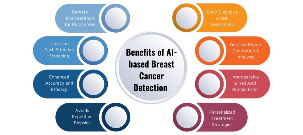
Benefits of AI reading mammograms
Conclusion
Artificial Intelligence in mammography has revolutionized diagnosis and prognosis of breast cancer by offering cost-effective solutions with enhanced accuracy and efficiency. In this blog, we discussed various ML algorithms, data processing methods and integration of these models with imaging modalities that enable a robust framework in order to develop AI-driven diagnostic tools. With time, challenges related to data quality, interpretability, and regulations are being addressed for clinical practices to fully realize the applications of AI and harness its benefits. Synergy between AI and radiomics holds immense potential and continuous collaboration between AI developers, researchers, clinicians, regulatory bodies and radiologists is crucial for improving diagnosis of breast cancer and better patient outcomes. KritiKal Solutions has worked with various clinical institutions and authorities for developing AI-based reporting solutions that allow radiologists to report and annotate images while providing image annotation services as well. The end-to-end mammography solutions enabled with segmentation and classification DL models, feature searchability within images and AI-based breast cancer prediction. Please mail us at sales@kritikalsolutions.com to know more and avail our Medtech services.
References:
- LeCun, Y., Bengio, Y., & Hinton, G. (2015). Deep learning. Nature, 521(7553), 436-444.
- Litjens, G., et al. (2017). A survey on deep learning in medical image analysis. Medical Image Analysis, 42, 60-88.
- Esteva, A., et al. (2017). Dermatologist-level classification of skin cancer with deep neural networks. Nature, 542(7639), 115-118.
- Aerts, H. J., et al. (2014). Decoding tumour phenotype by noninvasive imaging using a quantitative radiomics approach. Nature Communications, 5(1), 1-9.
- Rehman, A., et al. (2018). Deep learning in mammography and breast histology: Classification and detection of breast cancer. ACM Computing Surveys (CSUR), 51(1), 1-36.

Buddha Suchith currently holds the position of Senior Software Engineer at KritiKal Solutions. He has extensive experience in the Computer Vision domain and has been involved in multiple end to end computer vision based real time and POC projects using Python and C++ as main technologies. With his passion towards vision solutions and research, he has helped KritiKal in timely delivery of these projects to major SMBs and Fortune500 companies.