

 Global
  United States
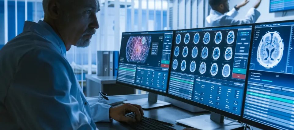
What is AI in Diagnostic Imaging?
Artificial Intelligence (AI) is continuously evolving across all sectors, including healthcare and medical technology, by accelerating interventions in foundational architectures of radiology and other types of diagnostic imaging. Combining the drivers, methodologies, interdisciplinary applications, and commercial products of AI with necessary points of medical imaging and disease management. Alongside the contemporary utilization of deep learning, advancements in multimodal models, and Generative Pre-trained Transformer (GPT) in medical technology solutions, the AI wave has been transforming various fields through driven investments and innovations. These may include research and academics, medicinal practices, traditional work structures, frameworks, and the radiological paradigm.
Diagnostic imaging refers to medical technologies that visualize body organs and other parts internally to monitor injuries and diseases. The radiology field utilizes X-rays, Computer Tomography (CT), Positron Emission Tomography (PET) scans, Magnetic Resonance Imaging (MRI), and ultrasound to perform related actions. Radiologists interpret and precisely analyze the images for guiding disease detection, surgical planning, and evaluating treatment decisions and effectiveness. Healthcare systems across the globe are facing increased patient loads, a surging number of diagnostic scans, and a shortage of radiologists. AI development services can bridge gaps related to operational efficiency and global access as well as offer precise and timely decision-making, especially in remote and underserved areas.
The market for artificial intelligence in medical imaging was valued at US $1.36 billion as of 2024 and is expected to surge at a CAGR of about 34.87% to reach an approximate value of US $19.78 billion by 2033. In this blog, we will discuss how businesses can harness the latent potential of AI, the development and integration of AI models in medicine and healthcare technology, advancements in disease mitigation and addressal at the intersection of diagnostics and technology – radiology and diagnostic imaging.
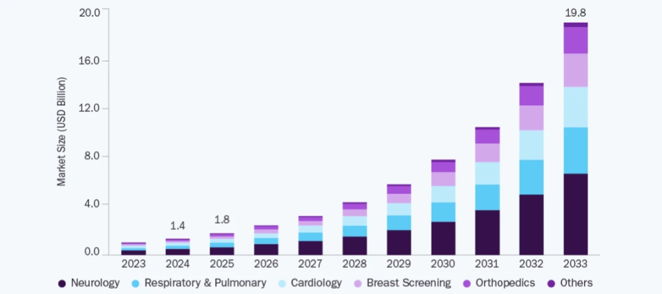
Growing market size of artificial intelligence in medical imaging during the 2023 to 2033 forecast period
How does AI in Medical Imaging Work?
Artificial intelligence is pushing the boundaries of image processing through the following measures.
Pattern Recognition
With the help of AI, radiologists can receive enhanced analysis, as these systems can identify complex patterns, especially in the case of specific or chronic diseases such as cancer, COVID-19, tuberculosis, and more. With swift, accurate, and early diagnosis, AI-powered image processing solutions can easily empower medical image interpretation and detect abnormalities in real-time. For example, AI can easily detect and triage pneumothorax or pleural effusion, which are the presence of air or fluid in the pleural cavity, respectively. Such conditions may be difficult to diagnose at first in an emergency setting where senior resources are constrained or short in number at a particular moment. Usage of AI for medical imaging results in effective patient management, emergency challenge addressal, regulation of high patient volumes, and overcoming the cons of limited resource availability all together.
Noise Reduction
Here, ‘noise’ refers to random variations in color-related information and brightness in radiological images, particularly in regions of light or shadow. It may originate from film grain in camera sensors and result in speckles, grainy textures, or banding patterns. AI through denoising autoencoders and Convolutional Neural Networks (CNNs) is employed in most radiological imaging environments to reduce electronic noise and enhance the clarity of low-light images effectively for accurate diagnoses. CNNs recognize key characteristics in the image, while denoising autoencoders organize necessary details for a clearer image.
Image Segmentation
Medical image analysis gets more precise and faster as AI outlines body organs, parts, pathological structures, components, and disease-affected regions in the obtained images. This assists radiologists in accelerating diagnosis, making informed treatment decisions, and forwarding the same to surgeons and doctors. For example, medical device contract manufacturing and AI systems development and service providers can introduce deep learning models like U-Net, which can be used to automate organ segmentation, lesion spotting, regional divisions, and disease tracking in the CT/MRI scans.
Abnormality Classification
Any kind of abnormalities, including tumors, fractures, infections like pneumonia, etc., can be easily detected and classified as per severity. For example, CNNs can identify COVID-19 symptoms, severity, and even make treatment-related predictions in imaging modalities like X-rays and MRIs. AI in diagnostic imaging helps radiologists in gaining a second opinion and performing comparable diagnostics within a short timeframe.
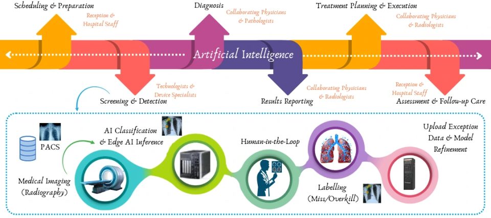
Sample workflow of integrating AI in diagnostic imaging
Predictive Modeling
As discussed earlier, AI solutions developed by medical device engineering services can analyze specific features in the images; at the same time, radiomics assists in extracting numerous quantitative features from the same. Machine learning models can therefore analyze large amounts of image characteristics. This aids in enabling prediction of disease progression, responses to treatment, patient outcomes, personalized treatment, and early detection of recurrences or malignancies for effective surgical planning.
Image Reconstruction
Deep learning-based denoising and super-resolution techniques can improve low-dose medical CT or fast MRI image quality. These image enhancement technologies can correct artifacts, reduce noise, and reconstruct high-resolution images from low-quality scans for a clearer visualization and precise diagnosis.
Triage Alerts
AI assists in streamlining and optimizing radiology workflows by prioritizing critical cases and automatically flagging urgent abnormalities. Triage tools developed through AI systems or wearable product design and development providers alert radiologists of high-risk findings by analyzing incoming scans. This reduces turnaround time and thus improves the functioning of high-volume settings like emergency departments for enhanced patient care.
Automated Reports
AI-powered Natural Language Processing (NLP) systems can analyze huge amounts of radiological reports to extract structured information and generate preliminary reports. Radiologists and involved specialists can link necessary imaging data findings with Electronic Health Records (EHR). This aids the management in facilitating data integration, maintaining consistency in reports, and supporting comprehensive clinical decisions.
Advantages of AI in Diagnostic Imaging
Here are some of the benefits of implementing AI in the medical imaging sector.
1. X-Ray: The software AI can detect pulmonary infections, bone fractures, thoracic diseases, cancer, sickle cell diseases, and lung abnormalities to avoid late or missed diagnoses in the initial assessments. It renders lower false positive rates and a higher curve for accuracy, even in rural areas.
2. MRI: AI identifies, enhances, and characterizes neurological disorders via automated segmentation and pattern recognition, including Alzheimer’s disease, musculoskeletal injuries, CT brain studies, strokes, multiple sclerosis, head injuries, etc. It reduces scan and evaluation time, optimizes imaging protocols, hastens diagnosis, and enhances patient comfort through standardized and detailed reports.
3. CT: It detects tumors, vascular diseases, midline shifts, intracranial hemorrhages, and skull fractures through real-time analysis of oncologic or neuroimaging and emergency cases, even in high volumes. Artificial intelligence in medical imaging covers deep learning algorithms that detect neurological conditions for error-free, faster, and accurate decision-making, especially in urgent head trauma cases as compared to fatigued or oversighted analysis.
4. Ultrasound: AI empowers automation of ultrasound-related techniques and key parametric measurements with precise fetal growth assessments, gestational age predictions, reproducibility and stability characteristics, abdominal organ analysis, and cardiac evaluations for minimizing user variability. AI-based models are especially useful for determining neonatal respiratory distress syndrome while improving accuracy, streamlining workflows, and enhancing clinical efficiency.
5. PET: Medical device design services can assist in integrating AI to improve quality and quantification of histopathological and radiological images for lesion and cancer detection. This helps in monitoring metabolic disorders, assessing patient risks, estimating survival results, and predicting treatment responses, even during shortages of specialists.
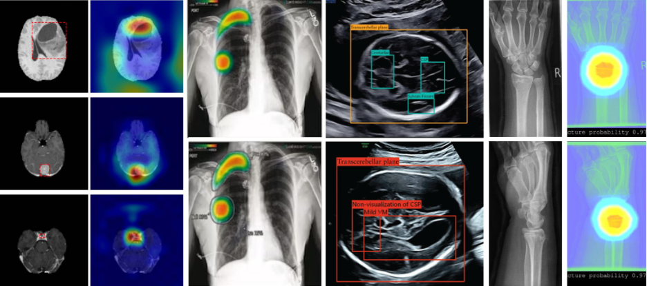
Various applications of artificial intelligence in medical imaging
KritiKal at the Forefront of AI in Medical Imaging
KritiKal’s specializations in the MedTech field range across various sectors and technologies, including AI model data annotation, training, testing, multi-platform optimization, porting, deployment, and real-time multi-camera video analysis. We can fine-tune existing generative AI, enterprise AI, and Large Language Models (LLMs) and integrate, deploy, and run them on premise servers. Our acumen in developing custom advanced enterprise software, cross-platform mobile applications, implementing Internet of Things (IoT) and Cloud-IoT infrastructure design and software engineering, visualizing and analyzing enterprise-scale data, designing, simulating, prototyping, and optimizing electronic circuits has driven multifold benefits for organizations.
With our command in product engineering, developing integrated solutions, tech stacks across technology domains, and proven track record of performing quality assurance checks, test automation, and compliances, a plethora of organizations have reached heights in terms of profits across the globe. Let us now delve into some of our AI-powered offerings specifically in the radiological imaging sector.
AI-powered Low Dose Imaging
We have enabled deep learning-enhanced low-dose imaging techniques for synthesizing full-dose quality images while preserving diagnostic accuracy for faster CT/MRI scanning workflows and enhanced patient comfort. Our solutions empower automated optimal alignment of patient positioning to reduce setup errors, preparation time, and artifacts for safer and higher-quality imaging via standardized practices in the case of vulnerable and frequent patients. We consider a plethora of metrics to track while applying AI for medical imaging, including structural similarity, lesion detection, signal, and contrast noise ratios, all built over a neural network-based encoder/decoder architecture.
Mammogram Tool for Breast Cancer Detection
Our offerings encompass an AI-powered automated tool for annotation, early detection, and reporting of breast cancer and related diseases via mammography to assist radiologists. It improves searchability within images for tackling cases of similar origin and features and saves lives. It showcases deep learning segmentation and classification models, image processing, AI prediction, and evaluation algorithms all packed into one, and it gathers continuous feedback from the operator for further improvements.
Vision-based X-Ray Image Stitching
Another important area that we have worked on is stitching full-body multiple X-ray images. The solution enhances images during preprocessing, aligns them via a vision-based algorithm using features from intensity and frequency domains, identifies stitch boundaries, compensates for exposure differences, and seamlessly blends the images. It therefore surpasses the limitations of full-body X-ray scanning techniques, detectors, and sensor limitations with C++ implementation for fine and fast image alignment. It reduces alignment errors through automated algorithms; it can assist in observing full-length leg images for surgical planning and is especially useful for diagnosis of scoliosis.
AI-powered Fetal Ultrasound Analyzer
We also offer AI models that automatically select and classify brain sub-planes into categories such as trans-cerebellum, trans-thalamic, and trans-ventricular. This assists ultrasound specialists in quick scans, accurate and detailed fetal anomaly ultrasound image viewing, analysis, diagnosis, and biometric measurement of brain-related features. This overcomes shortcomings of traditional methods such as operator dependency in manual selection and excessive time consumption.
Trained on the fetal head biometry dataset, the classification branch of the AI in medical imaging system can achieve unprecedented accuracy in head circumference measurement, that is, the perimeter of the fetal skull during the second and third trimesters. The AI models perform semantic segmentation and perimeter extraction using ellipse fitting in post-processing for consistent estimation of gestational age, monitoring of fetal growth, and detection of abnormalities.
3-D Guidance for Assisted Orthopedic Procedures
Apart from these, we offer a sophisticated 3-D guidance solution for orthopedic procedures that acts as a real-time fusion layer that overlays the CT map across the area of surgery. The edge-ready AI pipeline automatically registers live X-rays to the pre-operative CT volume. It combines scene-coordinate regression and PnP-RANSAC to compute rigid two-dimensional and three-dimensional alignment. All in all, our solution overcomes the cons of methods such as 2D C-arm fluoroscopy (X-ray), which is difficult to resolve depth and obscures anatomy while performing minimally invasive procedures.
It surpasses the shortcomings of traditional methods such as slow-paced manual alignment and clicks, error-prone procedures, increased exposure to radiation, and the requirement of fiducial markers. Trained in simulated Digitally Reconstructed Radiographs (DRRs), the solution features self-supervised domain adaptation in the operating room, offers robust and extreme views, occluding tools, and partial anatomy. Using OpenGL containerized edge deployment, it seamlessly integrates with existing hardware. It generates a 3-D overlay, which is updated on a continuous basis on the C-arm feed and easily streamed on any surgical navigation system or display. It uses a neural core registration engine for reduced operation room time and exposure to radiation. The extensive solution improves surgical accuracy and functions as per 3-D details related to changes in patient pose, tissue shift, and implants, once the incision is open, as compared to pre-operative CT image analysis.
In the above paragraph, PnP refers to Perspective-n-Point, a computer vision-based problem that seeks to determine the orientation, pose, and position of a given camera as per a set of ‘n’ 3D points in the world and their corresponding 2D projections in an image. It is widely used in augmented reality, robotics, and 3D reconstruction. RANSAC refers to a robust iterative method called Random Sample Consensus that estimates model parameters, including camera pose, etc., from a given dataset containing outliers. It samples data points in a random manner and fits a model continuously to search for the best-fitting consensus. Therefore, the RANSAC algorithm is used to solve the PnP problem, handle outliers in the 2D-3D correspondences, and improve accuracy.
Lung X-Ray Scanning for COVID & Pneumonia Detection
We developed an automated AI-based web application that guides doctors, pulmonologists, and radiologists to detect and assess the severity of COVID-19 cases during the pandemic. The assistive solution takes in lung X-ray images and makes accurate percentage-wise predictions regarding the possibility of COVID, pneumonia, and other lung-related diseases. The system features AI in medical imaging that performs swift image processing on the images, segments and classifies using deep learning models, and visualizes the results for clear assessment.
Conclusion: From Pixels to Precision in MedTech
Apart from the aforementioned proficiencies, KritiKal has assisted several large, mid-sized companies, startups, and even Fortune 500 organizations working in the healthcare technology sector to develop AI-powered medical imaging devices, technologies, platforms, and supporting software. Moreover, we showcase expertise in the design and development of various medical technology devices and AI-powered solutions and platforms, such as embryo grading software for IVF labs, explainable A-Fib detection through electrocardiogram (ECG) waveform analysis, polyp analysis colonoscopy or endoscopy for adenoma detection, cloud-based diagnostic fundus imaging devices, etc.
Businesses can leverage our decades of experience in the development of diagnostic medical devices for cardiovascular diseases, Gen AI-assisted infusion pumps, neuromuscular or electric muscle stimulation device, Wi-Fi-enabled gateways for patient support apparatus, wearable health monitoring devices, smart neckbands for monitoring the health of cattle and pets, and much more. We also offer agentic AI-based workflow for operational efficiency, document processing for insurance at hospitals, etc. We assure that all our solutions and products are secure, compliant, and developed by following adept cybersecurity practices.
We also bring to the table our acumen in the management of 510(k) submissions, risk management in medical device design and development with due adherence to regulations in healthcare such as ISO 14971, ISO 13485, European Union Medical Device Regulation (EU MDR), US Food and Drug Administration (US FDA) regulations, compliance practices for IEC 62304 for software lifecycle processes in medical technology, IEC 60601 standards for medical electrical safety, and more. We can perform threat modeling and devise risk mitigation strategies, integrate encryption and authentication mechanisms for safeguarding sensitive data, implement security controls as per FDA guidelines, and develop secure communication channels across Wi-Fi, Bluetooth, Universal Serial Bus (USB), and remote control. KritiKal has a proven track record in delivering high-security solutions in medical technology and conducting medical device testing like Vulnerability Assessment and Penetration Testing (VAPT) in phased approaches for developing medical devices.
Artificial intelligence in medical imaging is transforming the healthcare sector’s future with the help of continuous innovations in radiology and reshaping. AI-driven tools are enhancing the accuracy, speed, and efficiency of diagnostics right from the detection of abnormalities in CT, MRI scans, and X-rays to disease progression prediction. We understand critical issues associated with integrating AI in these setups, such as data privacy, security, model bias, and lack of interpretability, that currently hinder adoption. To tackle the same, our development practices accommodate fairness, transparency, and compatibility with existing infrastructure, thus ensuring trust and effectiveness of the solution. Please get in touch with us at sales@kritikalsolutions.com to know more about our solutions, products, and services and realize your MedTech requirements.

Ranbir Singh currently works as an Associate Software Engineer at KritiKal Solutions. He is proficiently skilled in full stack development, web development, working with ES6 features like Promises, Arrow function, HTML (5), CSS (3), MongoDB, React, Redux, Express, Next, JavaScript, Chakra UI, Bootstrap, Git, and more. With his ability to work efficiently in teams and strong expertise in developing AI/ML models, he has assisted KritiKal in delivering various projects to some major clients.